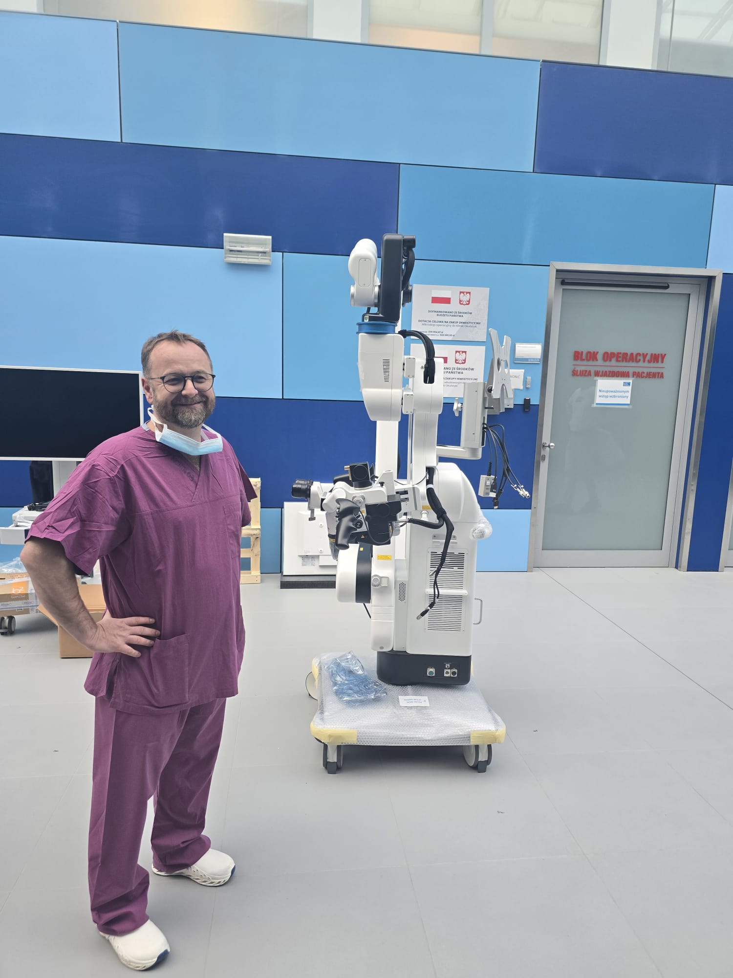
 One of the world's most modern neurosurgical microscopes was delivered to the MUB and University Clinical Hospital's Department of Neurosurgery before Christmas.
It will be used for high-precision brain and spinal cord surgery.
Nowadays, the microscope is no longer used only to view or magnify the operating field.
'It is now such a device that allows us to see much more and through various techniques,’ explains Tomasz Łysoń, MD, Head of the Department of Neurosurgery.
'It is now such a device that allows us to see much more and through various techniques,’ explains Tomasz Łysoń, MD, Head of the Department of Neurosurgery.
The new microscope gives enormous magnifications and perfectly illuminates the operating field. It was manufactured in Japan, by a company that specialises in designing lenses for telescopes and space satellites used for space exploration. It has an unusually high magnification capacity for objects, very fast zooming in and out. Just as importantly, it is equipped with various types of filters that allow surgeons to see much more than what they see with ordinary optics.
‘The filters with which the device is equipped enable us to depict a tumour, for example, very well,’ explains Dr Łysoń. 'We administer special drugs, which act fluorescently, and these become embedded in the tumour tissue. And then during the operation we have a special filter in the microscope, which allows us to depict only the tumour tissue. This diseased tissue simply glows a different colour. In addition to this, there are also special filters for viewing blood vessels, making it much easier to operate on various vascular pathologies.'
'Sometimes, when we operate on aneurysms or hemangiomas in the brain, we really have great difficulty in identifying, for example, what is an artery and what is a vein, because these vessels often look very similar,' adds Dr Łysoń. 'And this device will be very helpful.'
Neurosurgeons also enjoy another feature of the microscope - because it has the functions of an exoscope. That is, in addition to being able to look into the eyepiece, doctors can also look into a three-dimensional monitor. 'This is important in multi-hour surgeries, because then we don't have to bend over, focus, force ourselves into a head position, we just look at the monitor for ourselves during such a procedure,' explains doc Łysoń. ‘So it makes the work more comfortable and the surgeon does not get as tired, especially during longer procedures.
The market value of such a device is over PLN 3 million. The money for the purchase came from the state budget and was provided by the Ministry of Health.


